


The Science of Small
Top and cover photo by Rob Hess
This is an article from the spring 2015 issue of LSA Magazine. Read more stories from the magazine.
Labs in LSA are using Nobel Prize-winning microscope techniques to look closely at what scientists once thought would stay invisibly tiny—molecules moving around inside of cells. The new view magnifies some major possibilities for how we may one day see the world.
To get a sense of how small a bacterial cell is, picture a single grain of salt on a large armchair. Now imagine that you can stretch the size of the salt grain so it’s as big as the chair. A bacterium sitting on the grain of salt is about as tiny as the speck of salt relative to the armchair. In recent years, new methods of using microscopes have allowed researchers to see at even smaller scales than that—single molecules inside bacteria and other living cells.
The techniques won last year’s Nobel Prize in Chemistry for giving researchers a close-up view of what they once thought might be impossible to see: the real-time activity of individual molecules inside living cells. With these new approaches, LSA professors are gaining access to the tiny worlds of cells and relating those tiny worlds to the way our bodies work.
Sarah Veatch, an assistant professor of physics and biophysics, examines the interface between what’s inside a cell and what surrounds it—the cell membrane—using single-molecule tricks. Associate Professor of Cell and Developmental Biology and LSA alumna Kristen Verhey (’87) watches mobile molecules crawl along little tracks within a cell; she can even manipulate single molecules to measure the “horsepower” of their molecular motors. And Assistant Chemistry Professor Julie Biteen—who has worked with W. E. Moerner, one of the Nobel Prize winners—tracks the position of individual molecules as they go about their business inside a single tiny bacterial cell.
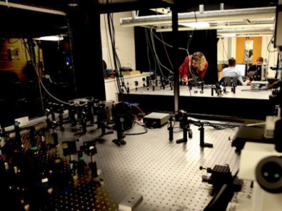
Assistant Professor Julie Biteen sets up lab experiments on optical tables in the lab. When she's ready to look under the microscope, she'll turn out the lights—the trick to single-molecule experiments is darkness, which gets rid of visual distractions in the cell. Lasers can then brighten the one molecule that she wants to see. Photo by Rob Hess
“What you’re looking at is this one protein that’s moving around through the cell, kind of hopping around,” Biteen says, referring to a movie she shot under a microscope of a bacterial protein called MutS. “And one thing we noticed is that it loiters for longer at the center. It’s moving in a sea of other proteins that haven’t found their targets yet. That distinction between one slow-moving population localized at the center and another faster-moving population that’s diffused all over the cell—a conventional experiment would really just wash out all those differences.”
While conventional test-tube experiments tend to stereotype similar molecules, crudely lumping them together as a group, single-molecule microscopy reveals just how unique individual molecules can be.
In Biteen’s experiment, for example, MutS proteins hang out in the bacterial cell, scanning DNA and searching for errors in the DNA code. Biteen is testing the hypothesis that MutS moves quickly and efficiently to correct errors in DNA. In a traditional experiment, she probably would have dumped her bacterial cells into a test tube, not knowing where MutS proteins traveled in the cell, nor how fast they moved. But single-molecule methods give an accurate, nuanced view of the interior of a bacterial cell.
“What we’ve uncovered is that, because of specific interactions with other proteins, the MutS proteins are poised and ready, so that they’re there when a mistake happens,” Biteen says. Dozens of MutS proteins constantly swimming around inside the cell thus act as “lifeguards” that save the cell from hazardous mistakes in the DNA code.
Single-molecule techniques allow researchers to note subtle differences among molecules, rather than rely on generalizations and assumptions based on how an average molecule tends to function in the cell. And the results potentially subvert our prejudices. For example, bacterial cells traditionally have had a reputation for being uncomplicated, primitive, gelatinous bags of stuff. As it turns out, they’re well ordered and rather sophisticated.
Now that we can see the impossibly small, it’s hard not to notice that the closer we look, the bigger our biases appear.
Lasers and Jellyfish
Looking at living cells—that’s a key advantage of single-molecule techniques. Although researchers have produced stunningly high-resolution images under a microscope by other means for some time, those methods required the specimens to be frozen, in a vacuum, or sliced razor-thin. In a word, dead. With single-molecule techniques, researchers can observe in high resolution the real-time activity and interactions among molecules in living cells. And these explorations are possible thanks to a jellyfish and a laser.
Researchers can attach fluorescent proteins that act both as labels and as little biological spotlights on the molecules they want to see under the microscope.
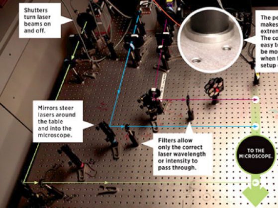
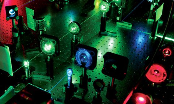
The wavelength of light determines the color of a laser beam. Different laser colors have different effects on molecules. Photo by Rob Hess
Fluorescent proteins glow brightly when shot by a laser, illuminating the position of each molecule. The first fluorescent protein came from a bioluminescent jellyfish. Since then, researchers have found other fluorescent proteins in various reef corals and have created synthetic versions in the lab. They’ve devised a series of ingenious methods of attaching the fluorescent labels to target molecules.
One of these methods involves inserting the fluorescent protein’s DNA into a cell’s DNA right next to, let’s say, the code for a target protein they want to examine. Each time the cell manufactures that protein by following the blueprint of its native DNA, the cell likewise creates a glowing label that’s integrated into the protein, based on the instructions in the introduced DNA. “The cell puts what I think of as a ‘backpack’ on each protein,” says Biteen.
This fluorescent backpack, conveniently manufactured and attached to the specific target protein by the cell’s own machinery, glows when it’s hit by a laser, allowing researchers to witness what goes on in a cell at the tiniest scale.
The Difference a Molecule Makes
Nils Walter has enormous ideas for what researchers can accomplish using single-molecule techniques. A professor in the Department of Chemistry, Walter directs LSA’s Single Molecule Analysis in Real-Time (SMART) Center, which he and an ad hoc think tank of other U-M researchers helped to establish in 2010 specifically to carry out single-molecule experiments. The SMART Center offers five microscopes, multicolored lasers, and personalized assistance in designing single-molecule experiments for anyone who asks.
“We are at the cusp of being able to actually look inside a live mouse brain,” Walter enthuses. In the past, researchers could employ fluorescent proteins to observe the mechanics of mouse cells, but the old methods gave researchers very little control over where those fluorescent labels would attach in a cell. But they’ve come a long way since then.
“Now, we have the tools to insert a fluorescent protein in a particular DNA or RNA sequence and label exactly this molecule in that position,” Walter says. By using the fluorescent backpack method in a living mouse, it’s possible to attach fluorescent proteins to the biological components that have relevance to a human disease and see exactly how those factors behave in a living system.
After that, it’s a matter of translating our understanding to human beings. “How we can really do what people refer to as ‘personalized medicine’ will depend on us understanding how these molecules actually interact, and how it’s different in different people,” Walter says. “We are individuals also at a molecular level.”
One day, a physician may be able to hold a device over a person to see exactly which of her molecules needs tweaking, so that a prescribed dosage of medicine can be fine-tuned—quickly and effectively—for each patient.
As methods in science penetrate increasingly small worlds, scientists can help to rapidly expand our perspective on how the larger world works. They accomplish all this by viewing particles so small that observing them closely was once thought impossible.
“People are doing this,” says Walter. “It’s not science fiction anymore.”