Please note: You may be required to obtain permission or training from the BSB Microscope Imaging Manager/Specialist before use.
The MCDB Imaging Labs contain advanced microscope equipment, image capture systems, software, and other resources available to all MCDB researchers and collaborators. This shared equipment is provided by the department to best aid as many research labs as possible.
Directions to the Imaging Suite
Take the east elevator. Scan your badge to select B to go down to the basement. Turn left out of the elevator and scan badge to access the microscope suite.
MCDB provides several research-grade microscopes with image capture systems to assist researchers with their sample imaging. [Room location in brackets.]
Confocal
Leica SP8 inverted laser scanning confocal microscope with 405nm and white light (470-670nm) laser systems, gating, spectral detection, resonant/tandem scanner, stage top incubator, and STED super resolution imaging with 592, 660, and 775nm depletion lines [B130]
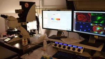
Leica Stellaris 5 inverted laser scanning confocal microscope with 405, 488, 561, and 638nm laser excitation lines available, spectral detection, image rotation, xz-y, and many other features [B138]
Leica/Andor Dragonfly Mosaic spinning disk confocal microscope imaging system with 405, 488, 561, and 638nm laser excitation lines, 10x, 20x, 60x, and 100x objective lenses, a FRAP module, photoconversion module, continuous focus feature, and dual Zyla cameras [4104C]
Leica SP5 upright laser scanning confocal microscope with 405, 458, 476, 488, 514, 561, and 633nm laser excitation lines available, spectral detection, and other features [5104H]
Nikon C2 inverted laser scanning confocal microscope with 405, 488, 543, and 638nm lasers. 20x and 60x objectives and stage top incubator [4104C]
Scientifica 2-photon confocal imaging system 1040nm laser, 10x and 16x LWD objective lenses
Florescence
Olympus IX81 inverted fluorescence microscope with motorized Z movement, a Retiga 1300R Fast, cooled, monochrome and color cameras, phase contrast, and 4x, 10x, 20x, and 40x objective lenses (60x dry and 60x oil available) [B138]
Leica MZ 16F fluorescence stereomicroscope with trans illumination base, 6MP color camera with fluorescence sensitivity, and microscope dichroics for UV, green (GFP), and red (RFP) fluorescence detection. [B110]
Nikon E800 fluorescence microscope with phase contrast, DIC, darkfield, UV, green (GFP), and red (RFP) LED excitation, a Nikon DS-Fi3 5.9MP color digital camera, and 4x, 10x, 20x, 40x, and 100x oil objective lenses. Provides color imaging with fluorescence sensitivity. [B110]
Leica Thunder fluorescence microscope with eight LED excitation lines, superfast large area tiling, and multi-area xy-z scanning, with instant computational clearing. 10x and 20x dry, 40x and 100x Oil objective lenses and incubation chamber. [B110]
TEM: Jeol JEM 1400plus Transmission Electron Microscope [B118]
Microscope Imaging Suite Specimen Preparation lab in B144A.
Leica 3040S cryostat sectioning, paraffin sectioning microtome station, Richiert Ultracut E ultra microtomes, paraffin and resin heating ovens.
If you need special specimen storage for use on the microscopes, please arrange with the Microscopy Suite Manager/Specialist.
Molecular Core: Access from the 3C LER.
Room 3215: StepOne Plus qPCR, 7500 Fast qPCR, CFX Connect qPCR, Nanodrop Lite, Qubit 2.0 fluorometer, Pipette Puller and evaluation microscope
Room 3235: Tecan SPARK Cyto plate reader, Tecan F200 plate reader, LiCor CLx IR imager, LiCor Fc UV and luminescence gel imager, GE Typhoon phosphor imager, Lyophilizer
Room 5235: Attune 4 laser flow cytometer, Fluorchem M UV and 3 color LED gel and mebrane imager

Before using any of these microscopes
Please speak with the Gregg Sobocinski, the BSB Microscope Imaging Manager/ Specialist.
- email: BSBimaging@umich.edu
- Phone: 734.615.2034
- Drop in: B140 BSB
If you have any questions about microscope technologies or accessories available within the MCDB department, please feel free to speak with the BSB Microscope Imaging Manager/Specialist, who will be happy to assist you with using these microscopes to their optimal capabilities or will recommend other University of Michigan resources, as appropriate.
Reserve Equipment
To reserve confocal or electron microscope equipment please use the iLabs MCDB Sign-up (link below). An active shortcode is required.
Instructions for iLabs login and use:
Internal User with a pre-created account