- All News
-
- Grad Student News
- Outreach News
- Search News
- Science Fun Facts
- Social Media
- Archived News
- Newsletters
- Expert Insights: Ben Hess on Importing Biological Materials
- U-M Herbarium Publication Spotlight: Dr. Thaís Vasconcelos and Dr. Aly Baumgartner Collaborate on Paper in New Phytologist
- U-M Museum of Zoology Publication Spotlight: Dr. Benjamin Winger's Study on Songbirds
- UMMZ Spotlight: Charlie Engelman Named to TIME’s "100 Most Influential Creators of 2025"
- Herbarium Spotlight: How AI is Transforming Specimen Transcription
- UMMZ Spotlight: A’liya Spinner is helping preserve the future of bees
- Meet the Researchers Driving Discovery Through the Biodiversity Exploration Fund
- EEB and U-M Museum of Natural History Celebrate ID Day
- All Events


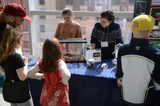
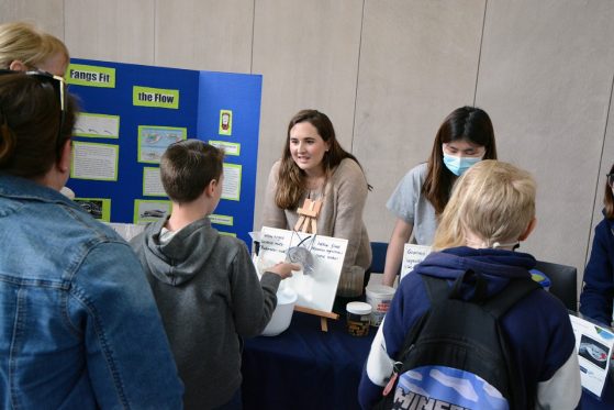
The event was a result of a partnership between the University of Michigan Museum of Natural History and Project MORPH!, which was produced by undergraduate students of EEB 450: Biology of Amphibians and Reptiles to translate herpetology into real-world applications that benefit both students and society in new ways.
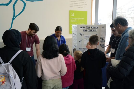
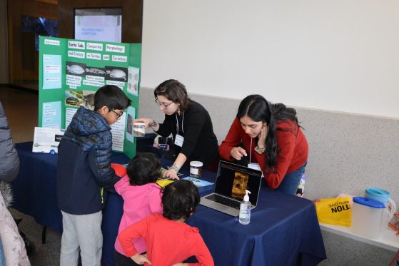
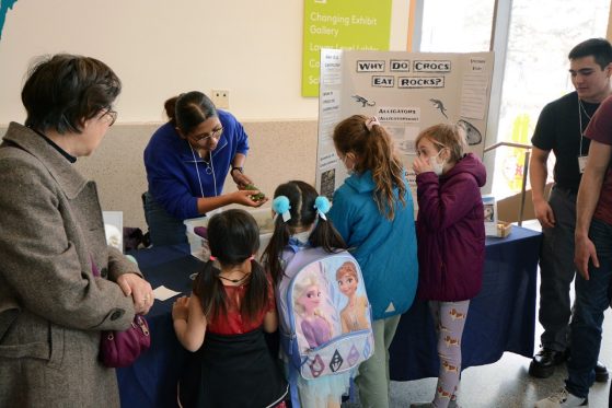
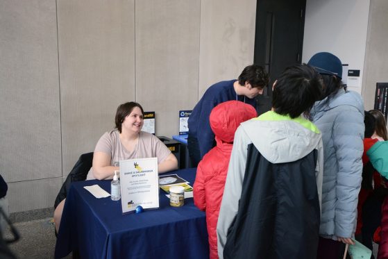
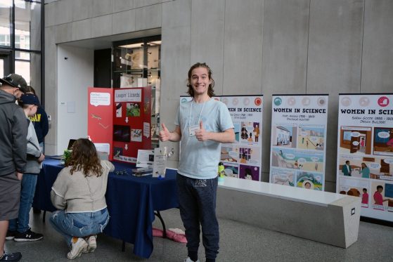
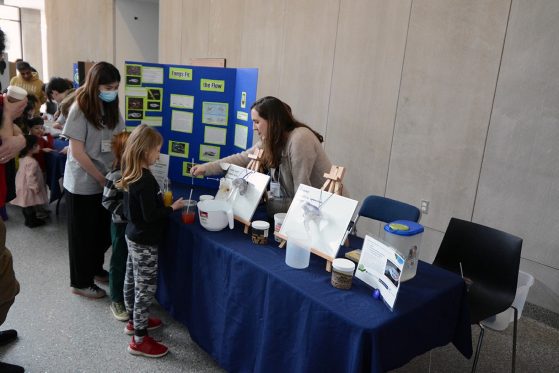
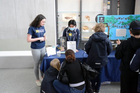
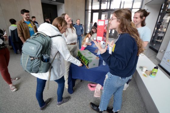
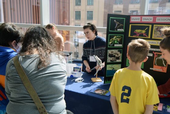
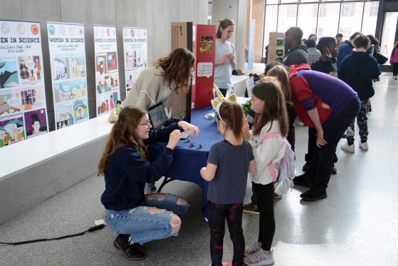
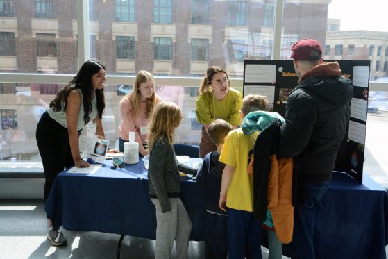
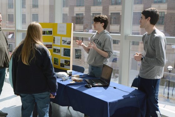
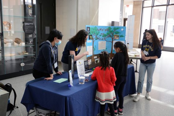
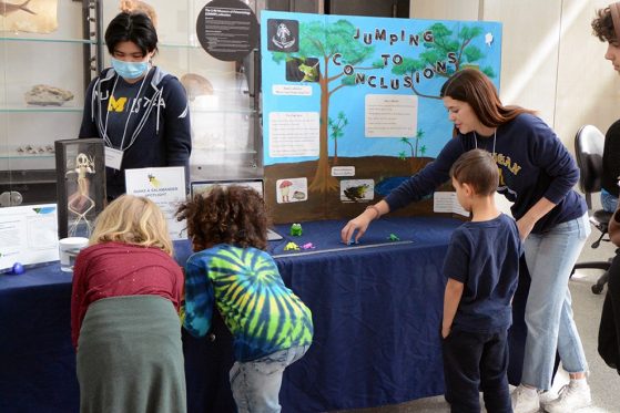
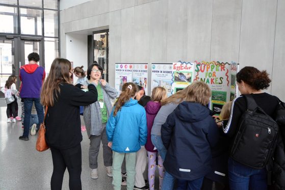
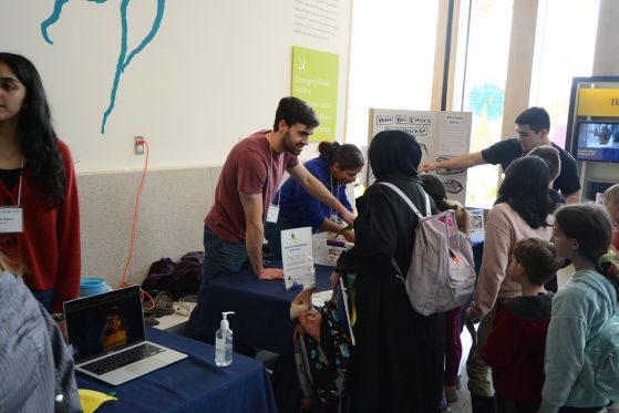
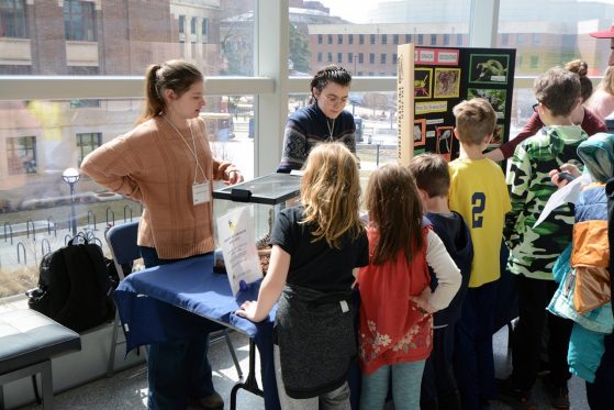
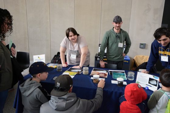
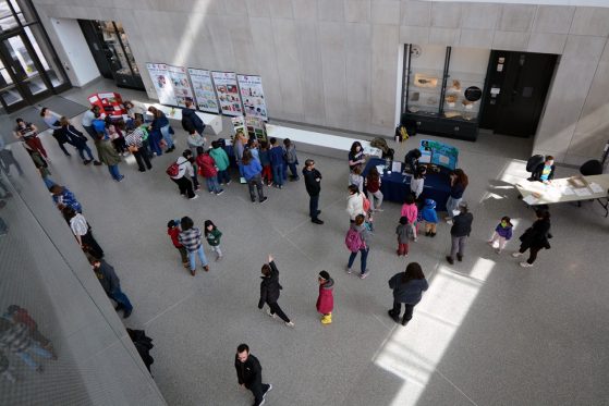
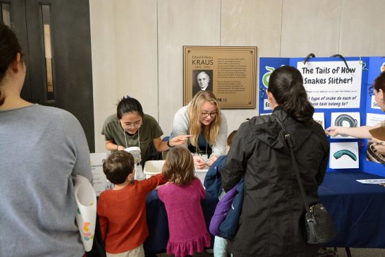
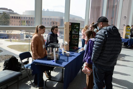
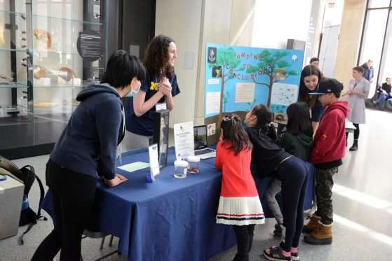
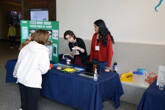
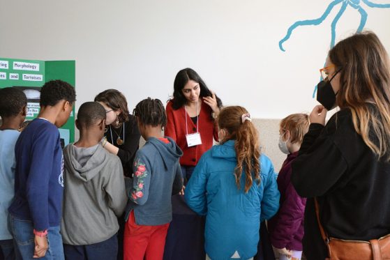
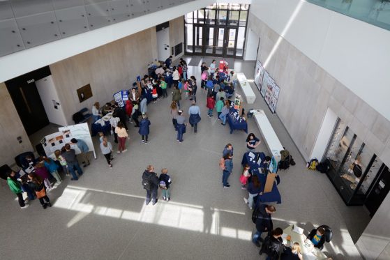
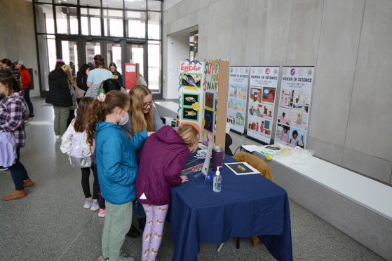
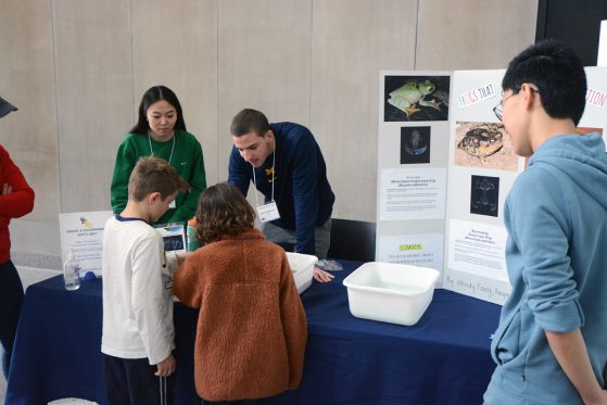
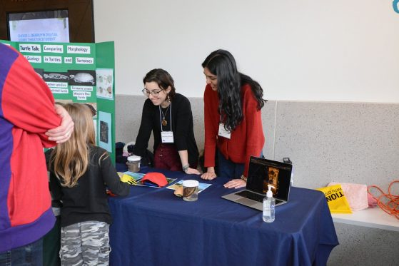
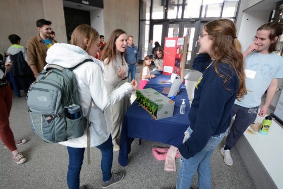
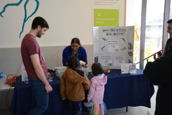
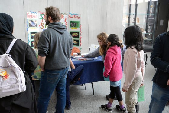
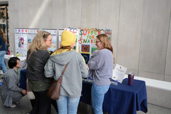
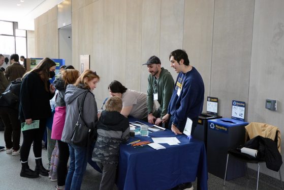
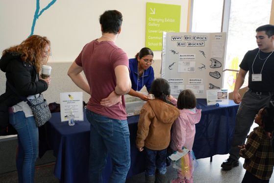
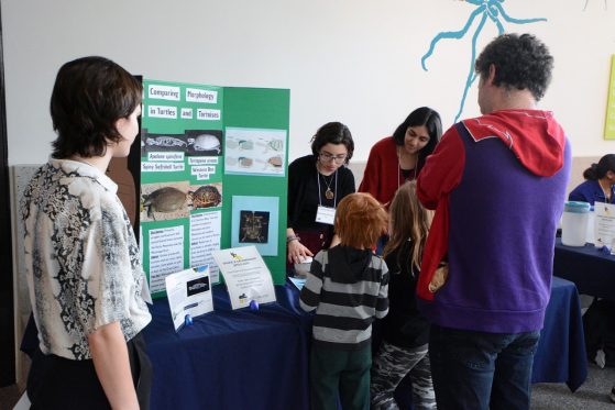
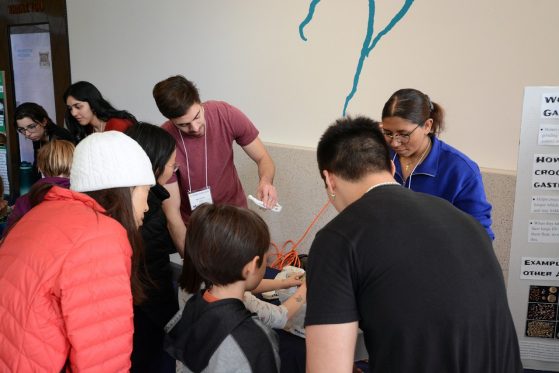
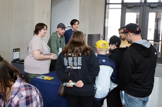
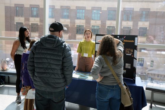
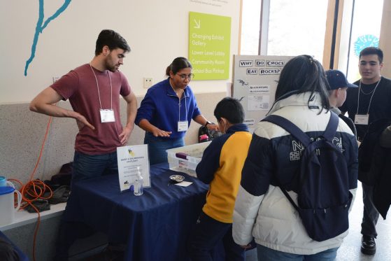
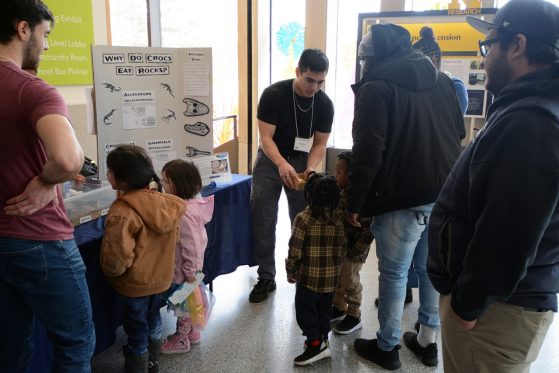
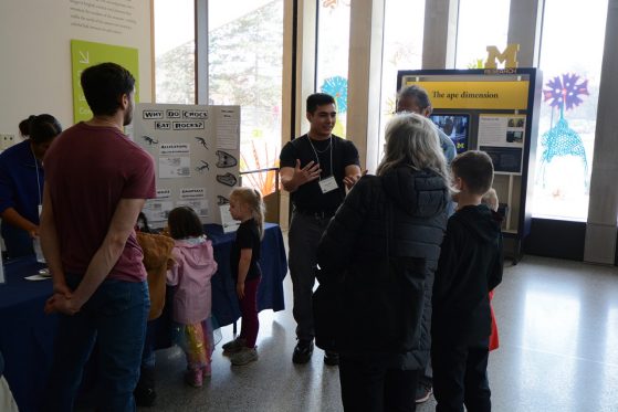
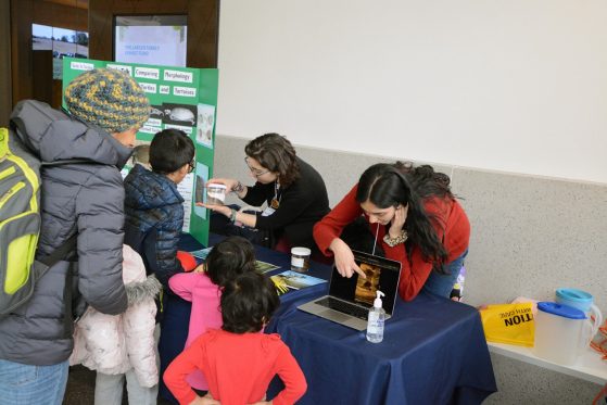
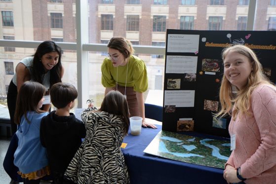
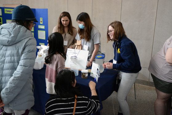
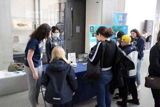
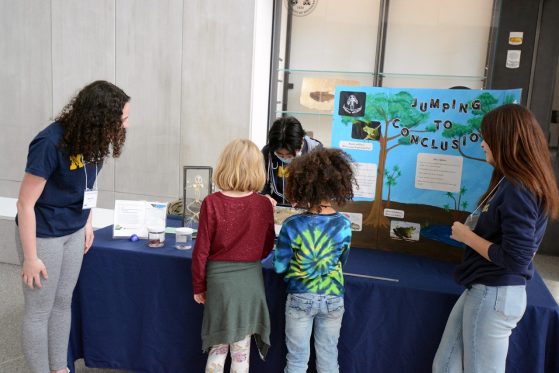
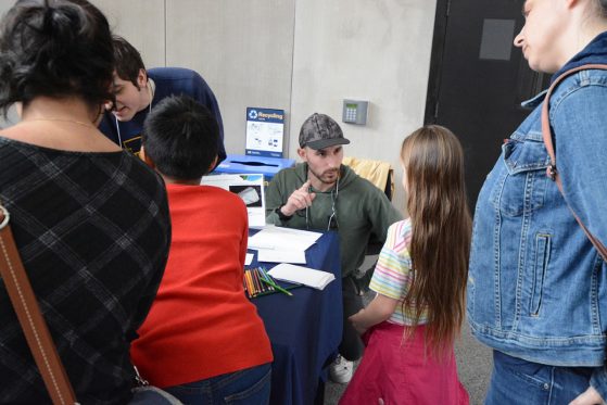
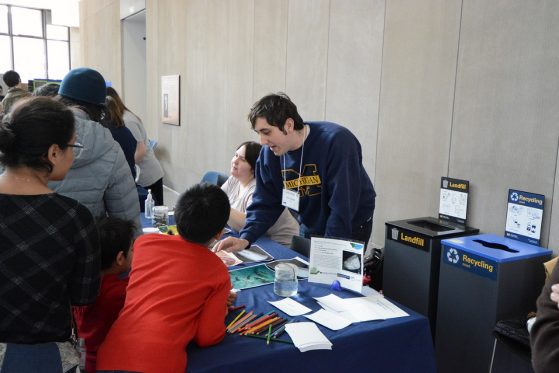
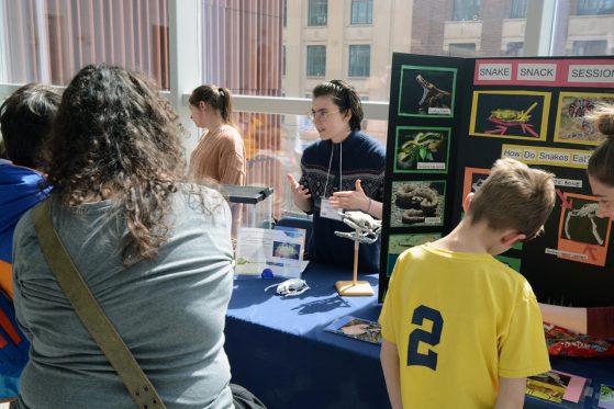
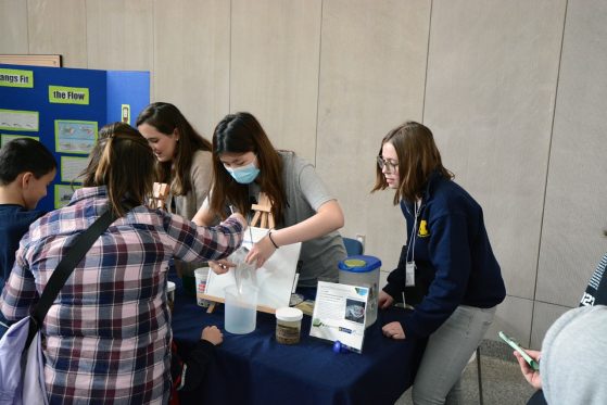
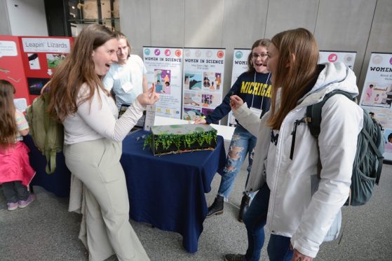
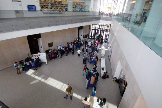
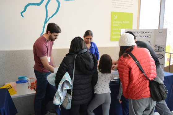
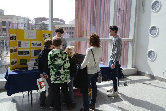
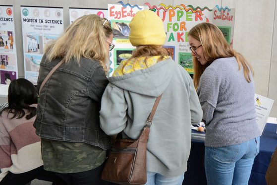
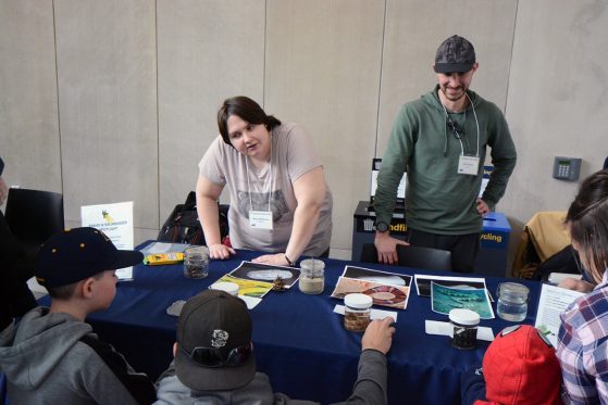
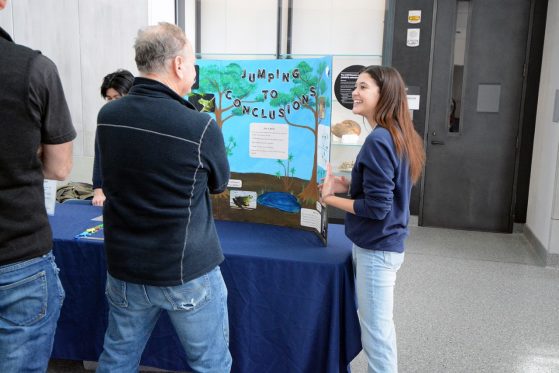
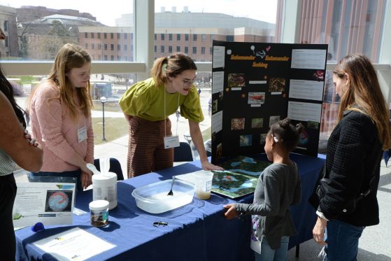
The Snake & Salamander event, led by Alison Davis Rabosky, assistant professor of ecology and evolutionary biology, welcomed visitors to the Biological Sciences building on March 28, 2023. Students from all over the Ann Arbor and Ypsilanti area came together during this spring break event to learn in a hands-on way. EEB 450 students engaged an all-ages crowd with hands-on explorations of amphibians and reptiles. This Biodiversity exhibit helped visitors understand the many unique features of reptiles and amphibians, from the iconic snakes and salamanders to the enigmatic caecilians, the limbless group of amphibians known as the “sharks of the soil.”
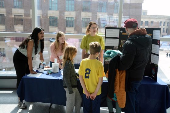
“The main goal of this event was to provide undergraduate students with the opportunity to share the knowledge they had learned over the course of the class while also improving their scientific communication skills. Additionally, they were also tasked with teaching the general public about how museum collections are used in scientific research,” said Hayley Crowell, EEB Ph.D. student.
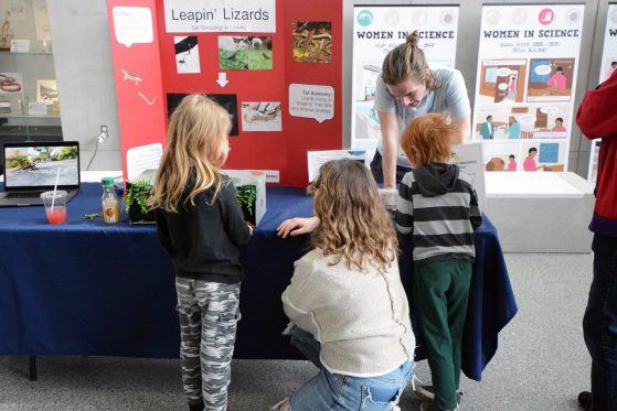
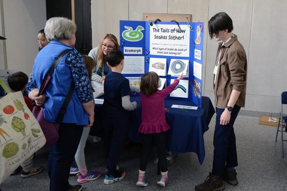
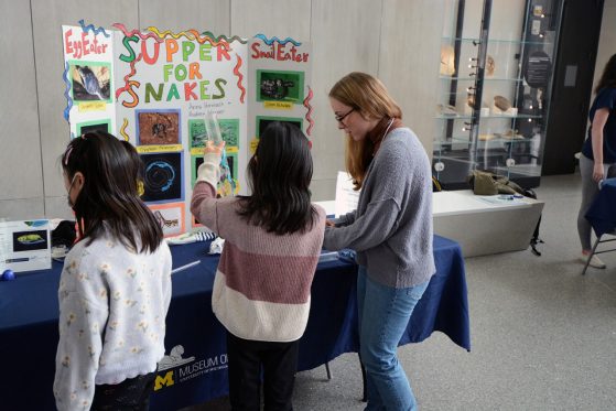
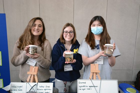
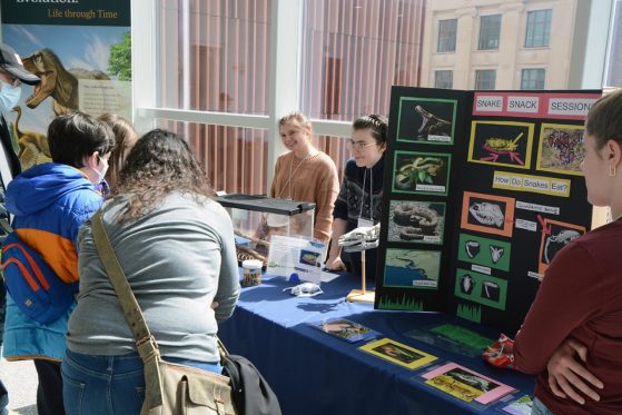
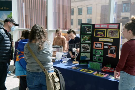
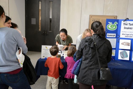
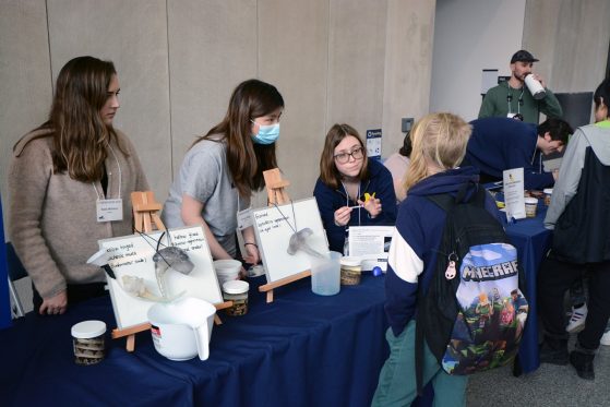
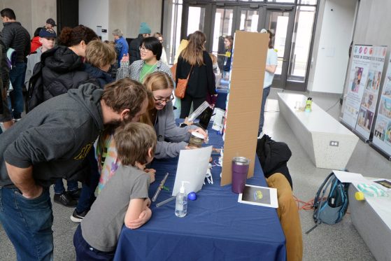
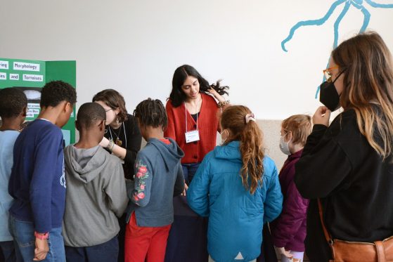
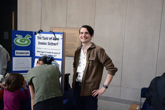
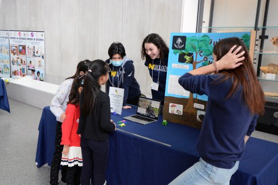
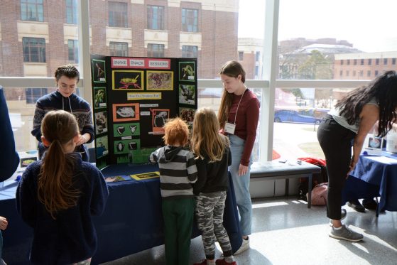
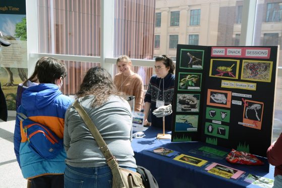
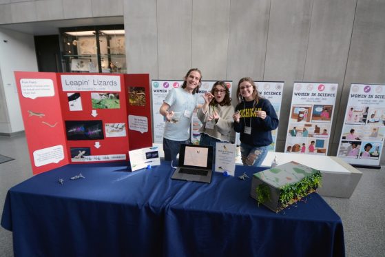
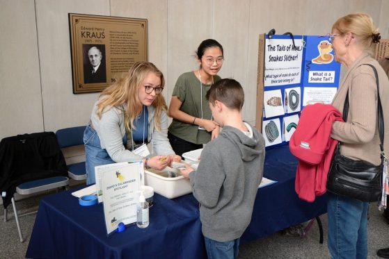
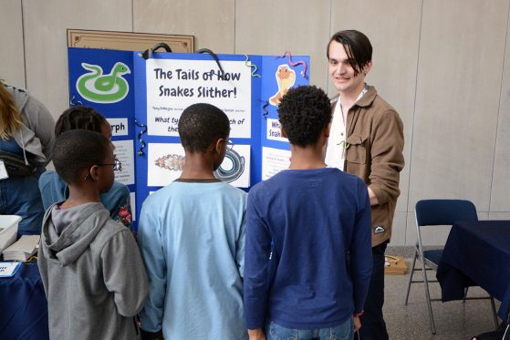
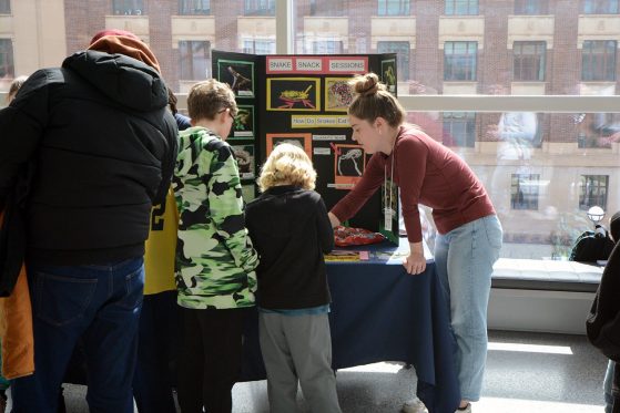
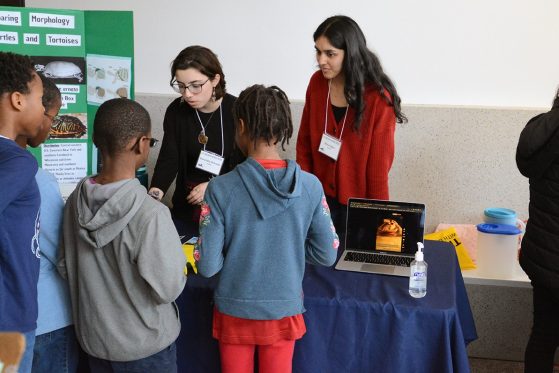
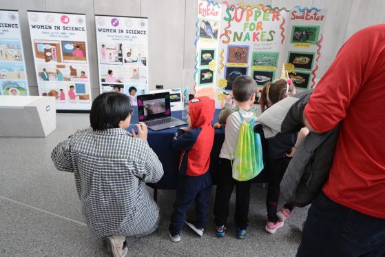
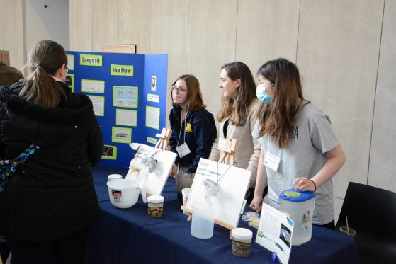
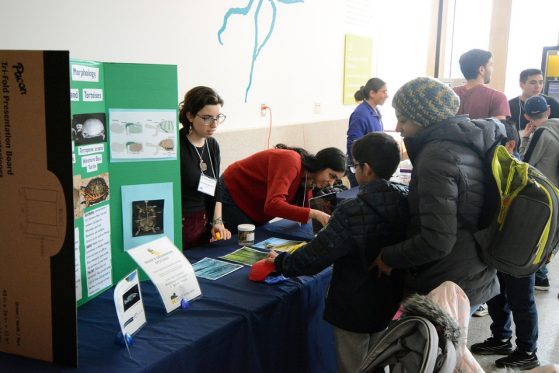
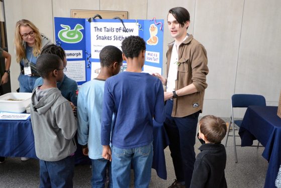
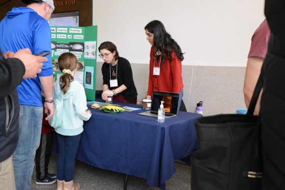
The collection of 12 different exhibits specifically featured the research of students who took EEB 450, who used 3D printers to help show the differences in snake tails for swimming versus climbing, illustrating how lizards can lose their tails when chased by a predator, and much more. Students used CT (computed tomography) scans of reptiles and amphibians created from real museum specimens. These scans help to communicate the importance of morphology in biodiversity science.
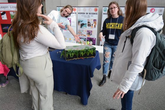
This event was part of Project: MORPH!, which aims to promote informal learning experiences by connecting students and community members. “We called it ‘Project: MORPH!,’ which was meant to signal the learning goals in all respects - that students translate their experience into a deeper understanding of morphological form and function, but also that both the students and public become “ambassadors” that help themselves and others value biodiversity in a compelling and memorable way,” said Davis Rabosky.
CT scans and other digital technologies are pushing the frontiers of biodiversity science by making specimens accessible in new ways. CT scans help to demystify specimens by providing a complex but accurate look of internal structures with more complexity than 2D images. Digital files and scans are more accessible around the world, opening possibilities for new discoveries. 3D printed models allow for scaling and, in turn, create a more hands-on experience as specimens enlarge. These 3D models also allow for up-close viewing of hidden structures. New technologies help to expand learning for students and community members alike.

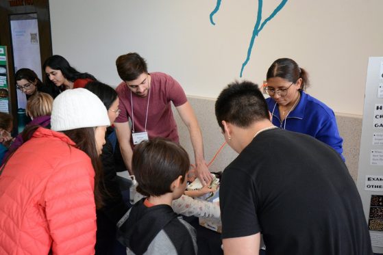
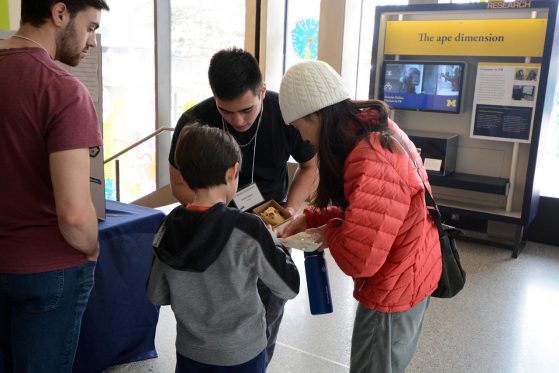
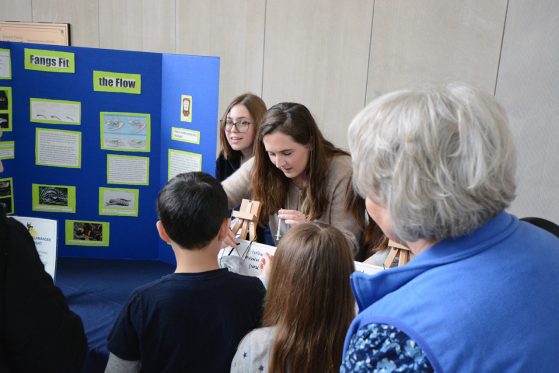
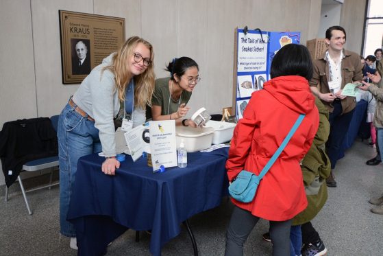
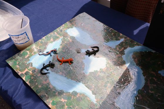
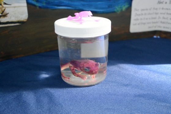
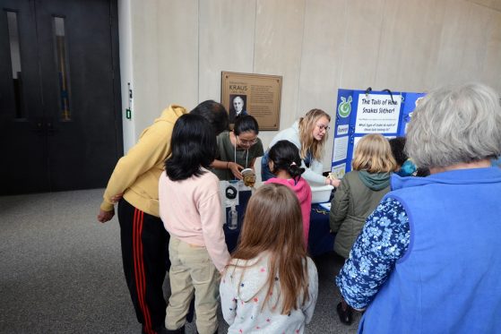
A special surprise was also revealed during a stop at Jada Lin, Tony DiMeglio, and Fern Sexton’s exhibit. These students used CT scans and visitor-guided 3D models in a sand and water table to show how snake tails work, with paddle-shaped tails making great sea snake swimmers. However, the CT scan also revealed that the snake [Shaw’s sea snake (Hydrophis curtus)] was pregnant. Another interesting feature of this scan is that the specimen was carrying live young instead of eggs, which is surprisingly common among species of lizards and snakes but not well appreciated. It’s likely collectors did not know this snake specimen was pregnant until these vital CT scans were created. With digital copies, this surprise can now be shared worldwide.
“My role in this museum event was to help organize the CT images and creation of the 3D prints. I was very excited to attend the outreach presentations and see how audiences received the final products; the enthusiasm by both students and museum visitors was impressive,” said Crowell. “Both children and adults alike were clearly enjoying the outreach activities and approached each table with a diverse plethora of biology questions.”
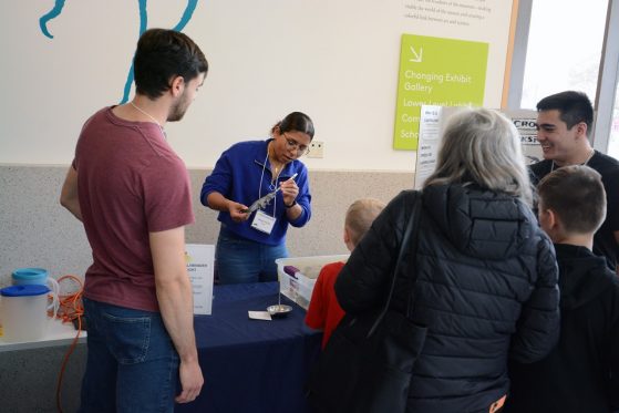
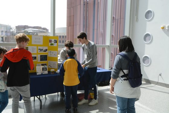
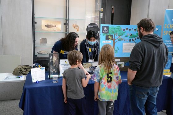
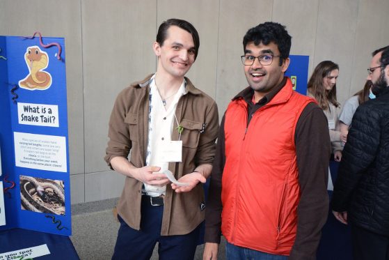
A broken tail gives clues to the Wright’s skink’s (Mabuya wrighti) ability to regrow tails. “This lizard specimen was collected from a private island and provides an excellent example of tail breaks (autotomy) in lizards,” writes students Aaron Blower, Alanha Rudd, and Fiona Corcoran. “It demonstrates how regrowable tails break in the middle of the vertebra (blue) rather than snapping in between the bones (in between blue and purple) as one might expect.” They connected audiences to this concept with a popular hands-on game that featured lizard models with detachable, magnetic tails, in which visitors played the role of hungry predators choosing prey items.
When asked about this event’s impact, Jade Marks, Science Communication Manager for the University of Michigan Museum of Natural History, highlighted the importance of students participating in this event. “There is a moment for the students when their science communication training, research, and prototyping coalesce and that moment is when they are face-to-face with a visitor who "gets it" for the first time,” said Marks. “It is incredibly rewarding to watch and it gives me hope for the future of our planet.”

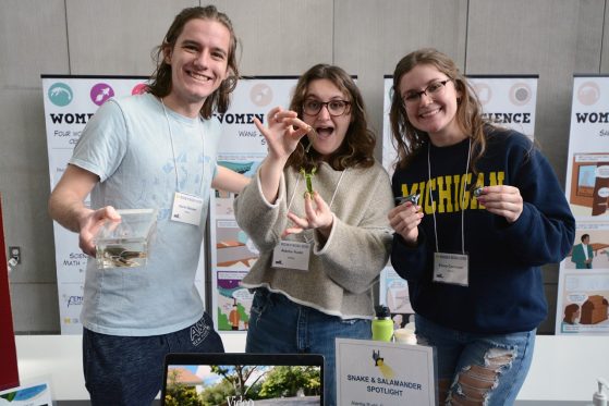
Thank you to Ecology and Evolutionary Biology graduate students Hayley Crowell, Natasha Stepanova, and John David Curlis, who assisted students with preparations, day of support, and photography. Thank you Ramon Nagesan, CT technician in the EEB Museums, Jade Marks, and Alicia Comer from UMMNH were integral to the success of this event. This event was also supported by the National Science Foundation (DEB-2141892 to Alison Davis Rabosky).